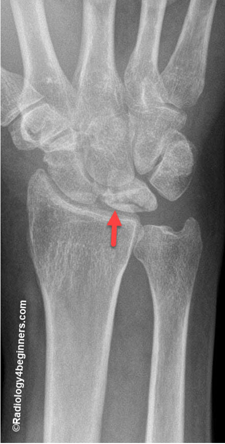
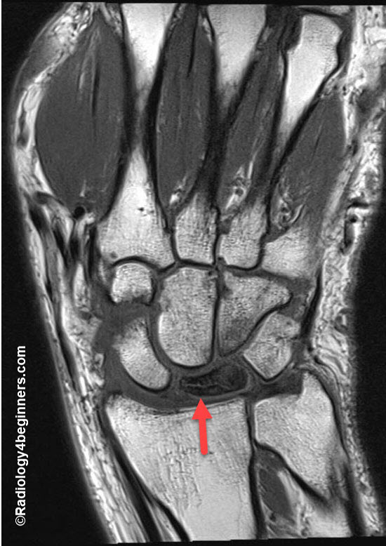
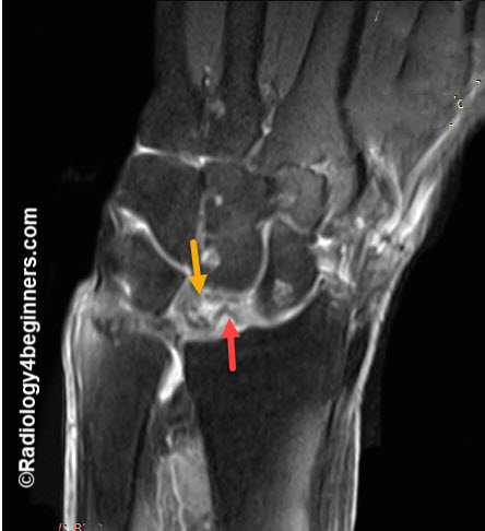
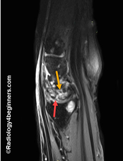
Stage I:
- May include a linear or compression fracture with normal lunate architecture and density.
- Pain is similar to a wrist sprain with mild activity-related wrist pain and pain with loading in wrist extension.
- Immobilization is widely used as therapy for Stage I disease.
Stage II:
- This stage shows increased density in the lunate on plain radiographs without a change in the shape, size, or relationship to other carpal bones. Often referred to as the first stage with visible changes on radiographs.
- Pain at this stage generally is more persistent. Treatment is still evolving. Joint-leveling procedures are often used.
Stage III:
- This stage is associated with pain both at rest and with activity.
- Stage IIIa: Fragmentation and deformation proximal(without fixed scaphoid rotation).
- Stage IIIb: Extended fragmentation (with fixed scaphoid rotation).
- This stage disease may be treated similar to Stage II with or without a revascularization procedure.
- Often treated with proximal row carpectomy or limited intercarpal fusion.
Stage IV
- Complete fragmentation
- Kienbock’s Disease Advanced Collapse (KDAC) to illustrate the parallels between Stage IV disease and scapholunate dissociation advanced collapse (SLAC) and scaphoid nonunion advanced collapse (SNAC).
- Treated with salvage procedures such as total wrist arthroplasty or total wrist arthrodesis.